
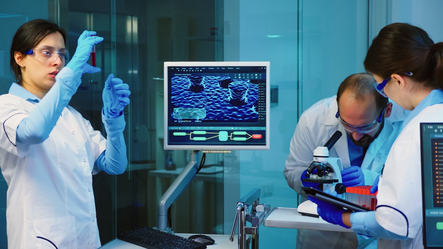
Precision matters when observation extends beyond what the human eye can process. A pathologist examining a tissue sample measuring 15x15mm faces an impossible choice: use low magnification to view the entire specimen whilst sacrificing cellular detail, or employ high magnification to see individual cells whilst losing spatial context. Manual stage manipulation introduces fatigue, positional inconsistency, and hours of tedious work that delays diagnosis.
Motorized XY stages solve this fundamental limitation by enabling automated scanning across large sample areas whilst maintaining micron-level positioning accuracy. These precision motion platforms integrate with optical microscopes to create automated imaging systems capable of capturing thousands of high-resolution images, stitching them into seamless panoramic views, and executing complex inspection protocols without human intervention. The technology transforms workflows across biomedical research, materials science, semiconductor manufacturing, and industrial quality control.
The Evolution of Automated Scanning in Biomedical Research
Motorized stages enable automated image stitching and large-area scanning essential for high-resolution panoramic imaging in pathology and pharmaceutical quality control by capturing hundreds of overlapping fields and computationally merging them into single continuous images spanning entire specimens. Digital pathology workflows depend critically on this capability, where whole slide imaging systems scan glass slides containing tissue biopsies at 20x or 40x magnification.
Traditional manual microscopy restricts pathologists to viewing small fields of view, typically 0.5-1.0mm diameter circles at diagnostic magnifications. A standard 15x15mm tissue section requires examining 225-900 separate fields to achieve complete coverage, consuming 30-60 minutes of continuous work. Human fatigue during extended sessions introduces inconsistency, whilst fragmented viewing makes spatial relationships difficult to appreciate.
Automated scanning with motorized stages completes the same task in 5-15 minutes whilst capturing every region with identical exposure, focus, and illumination conditions. The resulting panoramic images preserve spatial context, allowing pathologists to navigate seamlessly between overview and detail. Image analysis algorithms can then perform automated cell counting, measure staining intensity, or flag suspicious regions for expert review.
Pharmaceutical quality control laboratories employ similar workflows for filter paper inspection during sterility testing. Automated image stitching across entire filter surfaces reveals contamination patterns that spot-checking misses, improving detection sensitivity whilst reducing inspection time.
Precision Requirements for Microstructural Analysis in Metallurgy
Sub-micron resolution combined with positioning repeatability better than 2 micrometres allows consistent focus retention and positional accuracy when performing detailed grain structure analysis across large metallurgical specimen areas measuring 50-100mm in material science research. Metallurgical microscopy demands examination of polished and etched metal samples to reveal grain boundaries, phase distributions, and defect structures that determine mechanical properties.
Material scientists studying failure mechanisms in aerospace components need to correlate microstructural features with stress concentrations and crack initiation sites. This requires scanning large areas around failure locations whilst maintaining high magnification necessary to resolve grain-scale details. Manual stage positioning introduces coordinate uncertainties of 10-50 micrometres, making it impossible to return precisely to specific features for follow-up examination.
Motorized stages with digital coordinate systems enable researchers to record exact positions of features of interest, return to those locations days or weeks later with micron-level accuracy, and build comprehensive maps correlating microstructure with mechanical testing results. The ±5µm mechanical accuracy ensures that features identified in initial scans remain accurately positioned through subsequent examination cycles.
Large metallurgical samples often exceed 50x50mm in area. Motorized stages with 300x300mm travel range accommodate these specimens whilst maintaining positioning precision across the full working envelope. Vibration-free operation prevents image degradation during automated scanning, preserving image quality necessary for quantitative metallography measurements.
Semiconductor Inspection: Automating 300mm Wafer Analysis
Low defect tolerance in semiconductor manufacturing demands automated optical inspection (AOI) systems capable of detecting line breaks, shorts, and particle contamination across 300mm wafers, where motorized stages enable systematic scanning with 0.6 µm resolution to identify defects measuring just a few micrometres. Modern semiconductor fabrication operates at technology nodes below 10 nanometres, where microscopic defects cause device failures and yield losses.
Semiconductor manufacturers inspect wafers at multiple production stages: after photolithography to verify pattern transfer, following etching to confirm feature dimensions, and post-cleaning to detect particle contamination. Automated optical inspection systems integrate high-resolution microscopes with precision motorized stages to execute programmable scan patterns that systematically image entire wafer surfaces.
The stages position samples with repeatability measured in hundreds of nanometres, ensuring multiple inspection passes align accurately for before-after comparisons. Real-time image analysis algorithms running during scanning identify anomalies including line breaks, shorts, and particle contamination.
The economic impact is substantial. A single 300mm wafer may contain several hundred die valued at INR 5,000-50,000 each depending on device complexity. Early defect detection enables process corrections before entire batches become scrap, protecting investments potentially exceeding INR 50 lakh per wafer lot.
Beyond Imaging: Solar Cell Analysis and PCB Quality Control
Specialized applications including solar cell finger interruption detection and printed circuit board copper bridging inspection utilise high-travel motorized stages spanning 300x300mm to accommodate large samples whilst maintaining micron-level accuracy for defect identification. These industrial quality control applications demand different performance characteristics, prioritising large working envelopes and rapid scanning.
Solar cell manufacturing produces panels measuring 156x156mm or larger, where metallic grid fingers collect photogenerated current. Finger interruptions, breaks in these fine metal lines, reduce cell efficiency and power output. Automated inspection systems scan entire cell surfaces using motorized stages, employing image analysis to identify discontinuities requiring rework or rejection.
Printed circuit board inspection benefits from automated scanning, where solder joint quality, component placement accuracy, and copper trace integrity determine assembly reliability. Motorized stages enable systematic inspection of complex boards containing thousands of solder joints, identifying defects including insufficient solder, bridging between adjacent pads, and copper trace breaks.
Technical Checklist: Selecting a Motion Platform for High-Throughput Labs
Laboratories evaluating motion platforms must consider multiple technical and operational factors determining long-term performance and integration success.
|
Parameter |
Manual Stages |
Motorized Stages |
|
Positioning Speed |
5-20 mm/s |
1-750 mm/s |
|
Repeatability |
10-50 µm |
<0.5-2 µm |
|
Scan Coverage |
Limited by operator |
Complete automated coverage |
|
Software Integration |
None |
Full automation, data logging |
|
Throughput |
1-5 samples/hour |
10-60 samples/hour |
Key selection criteria include travel range matching maximum sample dimensions, positioning resolution appropriate to imaging magnification, and load capacity accommodating sample fixtures. Vibration-free operation becomes critical at magnifications above 20x where mechanical resonances degrade image quality.
Elevating Indian Scientific Engineering Standards
Motorized XY stages represent essential infrastructure supporting India’s advancement in biomedical research, semiconductor manufacturing, and precision industrial quality control.
As a trusted motorized stage manufacturer in India, Hexon delivers systems achieving 0.6 µm resolution with ±5µm mechanical accuracy across travel ranges up to 300x300mm, matching international performance standards whilst providing cost advantages through domestic manufacturing.
Hexon’s engineering capabilities address the complete spectrum of automated imaging requirements: compact benchtop systems for routine microscopy, industrial AOI platforms for semiconductor inspection, and custom configurations for specialised research applications. The noiseless motion control eliminates vibration artifacts, whilst intuitive software interfaces simplify workflow development.