

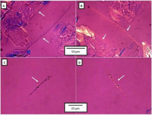
Asbestos analysis in talc is performed primarily through Polarized Light Microscopy (PLM), where analysts identify asbestos fibers by their unique optical properties rather than simply their appearance, following a structured seven-step workflow from sample preparation through USP-compliant reporting.
The analysis is primarily based on Polarized Light Microscopy (PLM), where the analyst identifies asbestos fibers by their unique optical properties rather than simply their appearance.
Step 1: Sample Preparation
A representative talc sample is dispersed in a refractive index liquid, mounted on a microscope slide, and cover-slipped, with multiple slides prepared to ensure a representative examination.
- A representative talc sample is collected.
- A small amount of talc is dispersed in an appropriate refractive index liquid.
- The sample is mounted on a microscope slide and covered with a cover slip.
- Multiple slides may be prepared to ensure representative examination.
Step 2: Initial Examination
Using the Olympus BX53 Polarized Light Microscope, the analyst screens the mounted sample under both plane-polarized and cross-polarized light, searching for elongated fibrous particles that visually resemble asbestos.
Using the Olympus BX53 Polarized Light Microscope:
- Observe under Plane Polarized Light (PPL).
- Observe under Cross Polarized Light (XPL).
- Search for elongated fibrous particles.
At this stage, the analyst is screening for fibers that visually resemble asbestos.
Step 3: Automated Sample Scanning
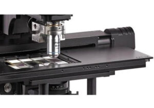
The Hexon Motorized XY Stage systematically scans the entire slide, recording coordinates of suspicious particles so large sample areas can be examined without missing regions and suspect fibers can be revisited automatically.
Using the Hexon Motorized XY Stage:
- The entire slide is scanned systematically.
- Coordinates of suspicious particles are recorded.
- Large sample areas can be examined without missing regions.
- Suspect fibers can be revisited automatically.
This significantly improves repeatability compared to manual stage movement.
Step 4: Optical Identification of Fibers
Each suspect fiber is evaluated for morphology and optical properties, then compared against the known optical signatures of six regulated asbestos minerals: chrysotile, amosite, crocidolite, tremolite, actinolite, and anthophyllite.
Each suspect fiber is evaluated for:
Morphology
- Length-to-width ratio
- Curved or straight fibers
- Fiber bundles
- Splitting characteristics
Optical Properties
The BX53 microscope allows measurement of:
- Refractive index
- Birefringence
- Pleochroism
- Extinction angle
- Sign of elongation
These properties are compared against known asbestos minerals:
- Chrysotile
- Amosite
- Crocidolite
- Tremolite
- Actinolite
- Anthophyllite
Step 5: Use of Compensators
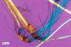
A first-order red compensator inserted into the optical path reveals fast and slow vibration directions and confirms sign of elongation, with the resulting colour changes under crossed polars serving as diagnostic evidence for asbestos mineral type.
A first-order red compensator is inserted into the optical path.
This helps determine:
- Fast and slow vibration directions
- Sign of elongation
- Confirmation of asbestos mineral type
The color changes observed under crossed polars are diagnostic for asbestos identification.
Step 6: Documentation
Every image, measurement, and observation is captured in Hexon’s CFR 21 Part 11 compliant software, with audit trails and electronic signatures making the record traceable and compliant with pharmaceutical quality systems.
Using Hexon’s CFR 21 Part 11 compliant software:
- Images are captured.
- Fiber measurements are stored.
- Analyst observations are recorded.
- Audit trails are maintained.
- Electronic signatures are applied.
Every observation becomes traceable and compliant with pharmaceutical quality systems.
Step 7: Reporting
The final report consolidates sample identification, microscopic observations, fiber images, optical characterization results, analyst conclusions, and electronic approval records into a single USP-compliant document.
The final report contains:
- Sample identification
- Microscopic observations
- Images of suspect fibers
- Optical characterization results
- Analyst conclusion
- Electronic approval records
Why the Motorized Stage Is Valuable
Because asbestos testing may require inspecting hundreds of microscopic fields per sample, the Hexon Motorized Stage’s automated mapping and coordinate-based relocation directly reduce operator fatigue while improving consistency and throughput.
For asbestos testing, laboratories may need to inspect hundreds of microscopic fields. The Hexon Motorized Stage enables:
- Automated slide mapping
- Large area scanning
- Coordinate-based relocation
- Reduced operator fatigue
- Improved consistency
- Faster USP <901>/<1901> workflows
Typical Workflow with Hexon Instruments
Talc Sample → Slide Preparation → BX53 Polarized Light Microscopy → Motorized Stage Automated Scanning → Fiber Identification → CFR 21 Part 11 Documentation → USP-Compliant Report
This combination of BX53 Polarized Light Microscopy, Hexon Motorized Stage, and CFR 21 Part 11 software provides a complete solution for asbestos screening and characterization in pharmaceutical and cosmetic talc according to USP guidelines.
FAQs
Q1. How many steps make up the USP <901>/<1901> asbestos testing procedure?
The procedure follows seven steps: sample preparation, initial examination, automated sample scanning, optical identification of fibers, use of compensators, documentation, and reporting.
Q2. What distinguishes an asbestos fiber from a look-alike particle under PLM?
Analysts rely on optical properties rather than appearance alone, evaluating refractive index, birefringence, pleochroism, extinction angle, and sign of elongation, then confirming with a first-order red compensator.
Q3. Which six minerals are asbestos fibers compared against during identification?
Suspect fibers are compared against the known optical properties of chrysotile, amosite, crocidolite, tremolite, actinolite, and anthophyllite.
Q4. Why is automated scanning preferred over manual stage movement?
The Hexon Motorized XY Stage scans the entire slide systematically, records coordinates of suspicious particles, and allows automatic revisiting of suspect fibers, which significantly improves repeatability compared to manual movement.